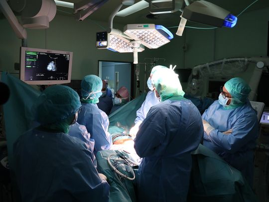
Last week at Latifa Hospital for Women and Children, a tiny little fetus, a brave mother and a team of highly specialised doctors created history when they performed the first-ever intrauterine fetal surgery for a spinal cord defect in the Arab region.
The extremely delicate six-hour surgery was performed on a 25 week old fetus that was diagnosed to have myelomeningocele, a form of spina bifida (spinal cord defect). The fetus weighed only 700 grams.
As the surgery was performed at this stage and not after the baby was born (which is typically still the standard medical procedure), the fetus’ defect was corrected, giving the baby a chance of improved cognitive function, better lower limb function and less risk of deformities.
Spina bifida is a neural tube defect that occurs during the first month of pregnancy when the spinal cord does not develop or close properly. In its most severe form, the defect leaves a section of the spinal cord and nerves exposed in a sac on the patient’s back.
Spina bifida can lead to many physical disabilities, including problems with walking and mobility, poor bowel and bladder function, issues with wounds healing and fluid accumulating in the brain (hydrocephalus), requiring shunt surgery.
DHA doctors highlighted this historic medical achievement for the Arab region at a press conference.
H.E. Humaid Al Qutami, Director General of the Dubai Health Authority said, “The DHA dedicates this great achievement to the wise leadership of the UAE, who always direct us to implement everything that achieves security, health and safety of community members.”
Multidisciplinary team
Dr Muna AbdulRazzaq Tahlak, Consultant in Obstetrics and Gynaecology and CEO of Latifa Women and Children Hospital, performed this surgery jointly with Dr Mohammad Sultan Al Olama, Consultant Neurosurgeon with Paediatrics and Functional Neurosurgery Specialty at Rashid Hospital and President of the Emirates Society of Neurological Surgeons in the UAE.
A highly specialised team of 20 multidisciplinary medical professionals from Latifa Women and Children Hospital and Rashid Hospital accompanied them during the surgery. The team comprised maternal and fetal medicine experts, obstetricians, anaesthesiologists, clinical pharmacologists, NICU nurses, scrub nurses from both hospitals, nurse coordinators and a radiographer.
Dr Tahlak said, “The patient was referred to us when she was 24 weeks pregnant after her condition was diagnosed in an ultrasound. We immediately conducted a full work-up. Upon analysis, the patient underwent comprehensive counselling to understand the situation at hand, the medical options and to prepare her for the surgery.”
The young 24 year-old Emirati patient, F.A., is a third time mum-to-be and has two healthy children. She said she wished to do everything possible for her baby’s well-being. “I want him to be a healthy boy and lead a healthy and happy life. As complicated and as difficult as the procedure was, I never hesitated even for a second about coming to Latifa Hospital and I am very thankful to the full team for everything they have done for my little boy and me.”
Precise surgery
Dr Tahlak said, “A multidisciplinary team was present for the surgery. This kind of surgery requires extreme precision and is a very delicate surgery. It requires opening the uterus but not in the way it is done for a C-section. In this particular case, the incision was done from the back of the uterus because her placenta was anterior. A minor incision was performed delicately – it had to be done in layers without opening the membrane that has the amniotic fluid and the fetus inside.
“We meticulously opened the membrane. We then used certain tools to extend the incision as the baby had a 6cm lesion in the spinal cord that needed to the fixed. We then gently positioned the baby in a way that the lesion was facing the incision so that Dr Mohammad Sultan Al Olama, the Neurosurgeon, could correct the spinal cord defect.”
Dr Al Olama said, “I first gave the fetus anaesthesia. Then, under the microscope, I started to repair the defect. There was an extra sac that I removed and then preserved the spinal cord and the nerves. Then I covered it delicately using micro instruments with layers of different membranes, sealed it and then covered that by the skin so that amniotic fluid does not touch the fetus’ spinal cord and this step is essential to prevent leakage of spinal fluid.
“I used 6.0 sutures to close this delicate defect. These sutures are considered one of the finest sutures used in surgery. It took me less than 50 minutes to correct the defect and achieve the normal anatomical form for the fetus.”

